
About Scoliosis
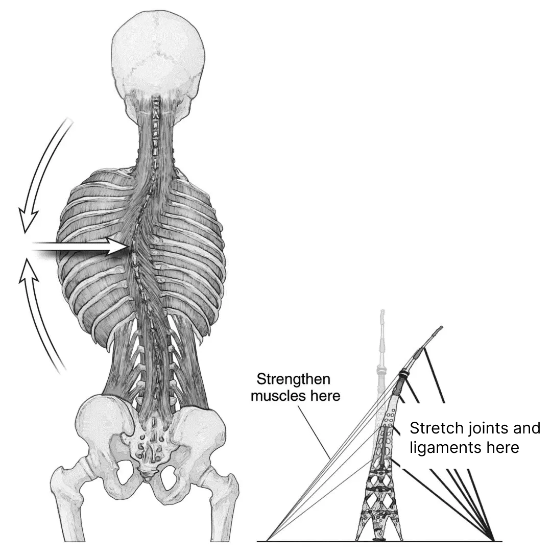
One major driver of juvenile idiopathic scoliosis is side-to-side muscle imbalance. Although we humans are largely symmetrical, there are always slight differences between one side and the other. The right thumbnail isn’t exactly like the left, one eye-tooth is a little bigger than the other and so on. Once in a while (2-3% of the time) the differences in the strength of the muscles on the right and left sides of the spine are large enough to tilt the spine toward the stronger side.
Consider a radio antenna held up by cables pulling down on the right and left, and suppose the cables on the right somehow were pulling down more powerfully. This imbalance would pull the antenna down to the right. This is exactly what frequently happens in idiopathic scoliosis.
For these reasons it’s best to work to correct scoliotic curves early in childhood: the muscular discrepancy between the two sides of the spine is smaller, and the resistance to straightening is weaker.
As time goes by, two things happen:
- Gravity gets ahold of the tilted spine and drags it down further, strretching and weakening the muscles on the left (convex) side.
- The ligaments and joints on the right (concave) side get closer together and knit their tissues tightly, making the spine more resistant to straightening out.
For these reasons it’s best to work to correct scoliotic curves early in childhood: the muscular discrepancy between the two sides of the spine is smaller, and the resistance to straightening is weaker.
For Medical Professionals
WHAT IS OUR METHOD, WHAT IS THE STUDY?
Our Approach:
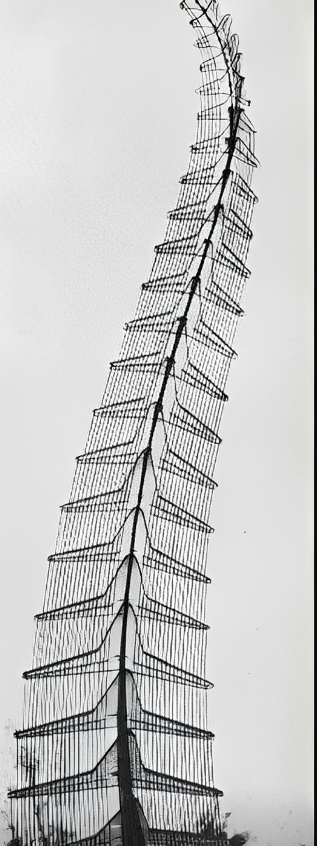
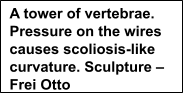
Background: We see the vertebral column as a tensegrity structure: one held up by tension created by the muscles that attach to it. To us, scoliosis seems to be due to asymmetry in the forces these muscles exert on different regions of the column. Those asymmetrical forces, generated by the muscles of the thoracic and lumbar spine, create and sustain the scoliosis. We have investigated this for more than 20 years. Using the simple yoga poses proposed here to strengthen muscles on the convex side of the curve, we have improved over 80% of the more than 300 patients we have treated2-7.
In this study we are testing the hypothesis that muscular imbalance is an important factor in generating and sustaining idiopathic scoliosis.
Approach:
Three factors motivate the proposed study:
- The spine can be seen as a tensegrity structure, one held together by tension between its parts. Asymmetry in that muscle-generated tension will curve the spine.
- We have encountered significant resistance from intra- and interarticular ligaments, facet joint capsules, vertebral asymmetries and asymmetrical fascial tightening that tend to preserve the status quo, i.e., the scoliotic curves. Being physically present in the operating theatre more than 100 times during scoliosis surgery, we have observed the significant resistance to surgically straightening the spine. The “soft tissues” harden, preserving the curve and resisting straightening these structures even with muscle-relaxing anaesthesia.
- In the course of this work, we have found that the people in earlier age groups tend to reverse their curves more quickly and more completely than older patients.
Currently the yoga, done daily to increase the strength and endurance capacity of convex-side muscles, reduces lumbar scoliotic curves by 2.5-3.5% per month2-7. We project that using isometric yoga poses in very young patients will not encounter the toughened resistance of ligaments, tendons and fascia that oppose the strengthening of the muscles of the convex side of the curve in older people, and thus will increase the efficacy of the treatment and shorten the treatment period. This is particularly important since the curves are naturally smaller at earlier periods in a person’s life and are easier to straighten on that account as well.
Study Design:
This is a two-armed randomized, blinded crossover study. After randomization each patient will receive a scoliosis X-ray. The intervention arm will do the intervention pose for 4 months, have another X-ray, and their part in the study will be over. The control group will do a placebo pose for 4 months, have another X-ray, then be taught the intervention pose and do it for 4 months and have a third X-ray. Then their participation will be terminated.
The Control Group pose that is good for the core, general strength and posture, but won’t help the scoliosis.
Radiation: Wherever possible, participants should have EOS X- rays, which have only 1/9th the radiation of the already low standard scoliosis X-rays. This means the radiation from the entire study will be no more than 1/3 of just one standard scoliosis X-ray. EOS technology is increasingly available in larger medical facilities such as hospitals, and in urban centers.
Statistical Analysis:
We will use mixed effects and MANCOVA and calculate Eta for effect strength.
Cost: The study is completely free and we will pay up to $100 for each X-ray when all X-ray studies are completed.
Study Objective Visit Schedule and Endpoints:
The objective of the study is to determine if judiciously applied yoga will expedite improvement in scoliosis, and to quantify any improvement detected. Cobb angle measurements, the long-time standard for measuring the size of scoliotic curves, will form the basis of the data.
The intervention group patients will have in toto two scoliosis films: X-rays: 1) within 3 months prior to teaching, or within 72 hours afterwards, 2) Four months following teaching. Each control group patient will have in toto three scoliosis films: X-rays: 1) within 3 months prior to teaching, or within 72 hours afterwards, 2) four months following teaching the placebo protocol, 3) four months after beginning intervention protocol. Although there are no required subsequent visits the study staff is available to counsel the patients and their families at any time and to help sustain the young patients’ efforts. Patients and their consenting families will be notified of the results of all X-ray studies, of course.
The endpoint of the study is when the last Control Group patient has had her or his final X-ray, approximately eight months after entering the study. Intervention group patients will be in the study only 4 months.